Optical imaging with high-end microscopy is a core element in CitBIO and essential towards our goals of achieving mechanistic understanding of intestinal peptide transport. This WP performs single-peptide tracking with state-of-the-art spatial and temporal resolution. To facilitate this, CitBIO, in collaboration with the Department of Health Technology, DTU, is establishing a unique microscopy facility with three newly acquired/custom-built microscopes:
- Nikon Ti2-E Yokogawa spinning disc confocal microscope.
- Custom-built two-photon laser-scanning microscope for large-volume tissue imaging.
- Custom-built lattice light-sheet microscope with adaptive optics for ultra high-resolution imaging of cells, intact tissues, and tissue models.
All microscopes are optimized for live-cell imaging. To facilitate research, this WP interfaces with all initiatives in the center, including our external partners. In particular, we work closely with WP3 and WP4 in order to develop new sample- and imaging solutions for the complex in vitro models developed in the center. Finally, we have a constant interplay with WP6 for development of novel data analysis tools for extracting quantitative insights from microscopy data.
WP members
Niels Bent Larsen, Professor
Rodolphe Marie, Associate Professor
Emil Boye Kromann, Associate Professor
Jannik Bruun Larsen, Post Doc
Kim Mortensen, Senior Researcher
Adam Hundahl, PhD student
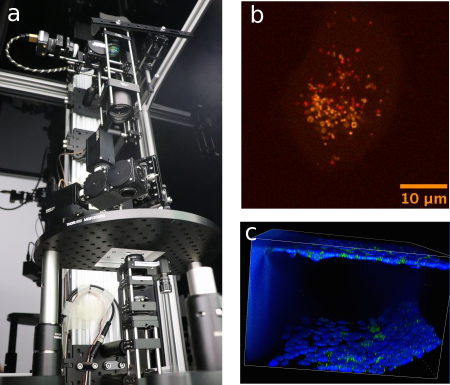
Figure: (a) Custom-built two-photon laser-scanning microscope for large-volume tissue imaging. (b) 4D Live-cell imaging of intra-cellular particles, structures, and organelles using spinning disk confocal microscopy. (c) 3D imaging of Caco-2 cell layers in a microfluidic chip using spinning disk confocal microscopy.